on September 11, 2023, 5:12 AM
NCERT Solutions for Class 11 Biology Chapter 6 Anatomy of Flowering Plants in Hindi and English Medium to Study online or download in PDF format free for new academic session 2024-25. Download NCERT Books in updated format for the current session 2024-25. Join Discussion Forum to share your knowledge with the others.
NCERT Solutions for Class 11 Biology Chapter 6
| Class: 11 | Biology |
| Chapter 6: | Anatomy of Flowering Plants |
| Content: | Exercises Solutions and Study Material |
| Content Mode: | Images, PDF, Text and Video Format |
| Academic Session: | Year 2024-25 |
| Medium: | Hindi and English Medium |
Class 11 Biology Chapter 6 Solutions in English
NCERT Solutions for Class 11 Biology Chapter 6 in PDF form to free download for new session 2024-25. Download NCERT Books 2024-25 based on latest CBSE Syllabus for new session. Offline Apps and NCERT Solutions are also available to free download. Discussion Forum is a platform to share your knowledge with each other.
Solutions App for Class 11


Important Terms on Anatomy of Flowering Plants
1. Axillary bud: The buds which are present in the axils of leaves (Consist of cells left behind from shoot apical meristem) and are responsible for forming branches of flowers.
2. Permanent tissues: The permanent tissues are derived from meristematic tissue, are composed of cells, which have lost the ability to divide and have become structurally and functionally specialised.
ANATOMY
Anatomy is the study of internal structure of organisms. Plant anatomy includes organisation and structure of tissues. Tissue is a group is cells having a common origin and usually performing a common function. Its wall is made up of cellulose. It performs the functions like photosynthesis, storage, secretion.
Collenchyma
Collenchyma: It is formed of living, closely packed cells. Its cells are thicknened at the corners due to depositon of celluose and pectin. It provide mechanical support to the growing parts of the plant. It is either found in homogenous layer or patches.
Sclerenchyma: It is formed of dead cells with thick and lignified walls. Provide mechanical support to organs. They have two types of cells: fibres and sclereids.
Xylem
I. Xylem consists of tracheids vessels, xylem fibres and xylem parenchyma. It conducts water and minerals from roots to other parts of plant.
1. Tracheids-Tube like cells with thick and lignified walls and tapering ends; dead, without protoplasm.
2. Vessel-long cylindrical structure made up of many cells with large central cavity, devoid of protoplasm. Present in angiosperms.
3. Xylem fibres-highly thickened walls; with obliterated lumens; septate or aseptate.
4. Xylem parenchyma-living and thin walled; cell walls made up cellulose, store food material in form of starch or fat. Radial conduction of water takes place by ray parenchymatous cells.
II. Protoxylem: The first formed primary xylem elements.
III. Metaxylem: The later formed primary xylem.
IV. Endarch: Protoxylem lies towards the centre and metaxylem towards the periphery of the organ; in stem.
Phloem
I. Phloem consists of sieve tube elements, companion cells, phloem fibres and phloem parenchyma; 1. Phloem transports the food material from leaves to various parts of the plant.
Sieve tube elements: These are long tube like structures arranged longitudinally, associated with companion cells. Its end walls are perforated to form sieve plates and functions of sieve tubes are controlled by the nucleus of companion cells.
2. Companion cells are specialised parenchymatous cells associated with sieve tube elements and connected with sieve tube elements by pit fields present between their common longitudinal walls. It help maintain pressure gradient in sieve tubes.
3. Phloem Parenchyma is made up of elongated, tapering cylindrical cells with dense cytoplasm and nucleus. Its cell wall made of cellulose with pits through which plasmodesmatal connections exist between cells and store food material.
4. Phloem fibers (bast fibers) are sclerenchymatous; absent in primary phloem but present in secondary phloem and elongated, unbranched pointed, needle like apices with thick cell walls.
II. Protophloem: First formed phloem with narrow sieve tubes.
III. Metaphloem: Later formed phloem with bigger sieve tubes.
Important Questions on 11th Biology Chapter 6
State the location and function of different types of meristem.
Meristems are specialised regions of plant growth. The meristems mark the regions where active cell division and rapid division of cells take place. Meristems are of three types depending on their location. Apical meristem It is present at the root apex and the shoot apex. The shoot apical meristem is present at the tip of the shoots and its active division results in the elongation of the stem and formation of new leaves. The root apical meristem helps in root elongation. Intercalary meristem It is present between the masses of mature tissues present at the bases of the leaves of grasses. It helps in the regeneration of grasses after they have been grazed by herbivores. Since the intercalary meristem and the apical meristem appear early in a plant’s life, they constitute the primary meristem. Lateral meristem It appears in the mature tissues of roots and shoots. It is called the secondary meristem as it appears later in a plant’s life. It helps in adding secondary tissues to the plant body and in increasing the girth of plants. Examples include fascicular cambium, interfascicular cambium, and cork cambium.
Cork cambium forms tissues that form the cork. Do you agree with this statement?
When secondary growth occurs in the dicot stem and root, the epidermal layer gets broken. There is a need to replace the outer epidermal cells for providing protection to the stem and root from infections. Therefore, the cork cambium develops from the cortical region. It is also known as phellogen and is composed of thin-walled rectangular cells. It cuts off cells toward both sides. The cells on the outer side get differentiated into the cork or phellem, while the cells on the inside give rise to the secondary cortex or phelloderm. The cork is impervious to water, but allows gaseous exchange through the lenticels. Phellogen, phellem, and phelloderm together constitute the periderm.
Explain the process of secondary growth in stems of woody angiosperm. What is the significance?
In woody dicots, the strip of cambium present between the primary xylem and phloem is called the interfascicular cambium. The interfascicular cambium is formed from the cells of the medullary rays adjoining the interfascicular cambium. This results in the formation of a continuous cambium ring. The cambium cuts off new cells toward its either sides. The cells present toward the outside differentiate into the secondary phloem, while the cells cut off toward the pith give rise to the secondary xylem. The amount of the secondary xylem produced is more than that of the secondary phloem. The secondary growth in plants increases the girth of plants, increases the amount of water and nutrients to support the growing number of leaves, and also provides support to plants.
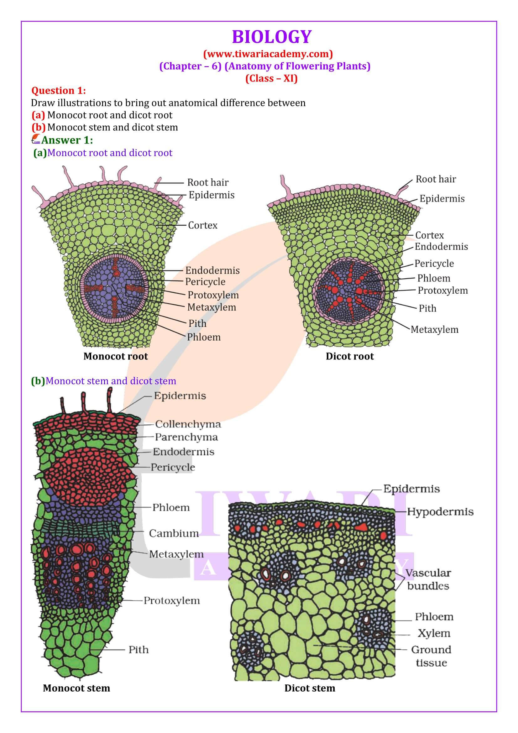
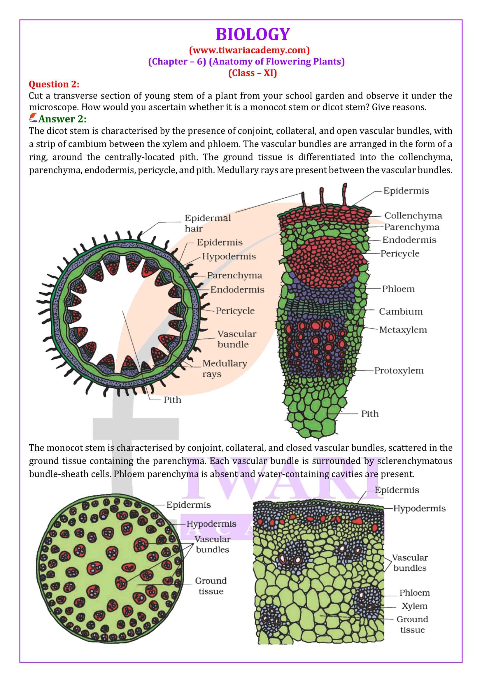
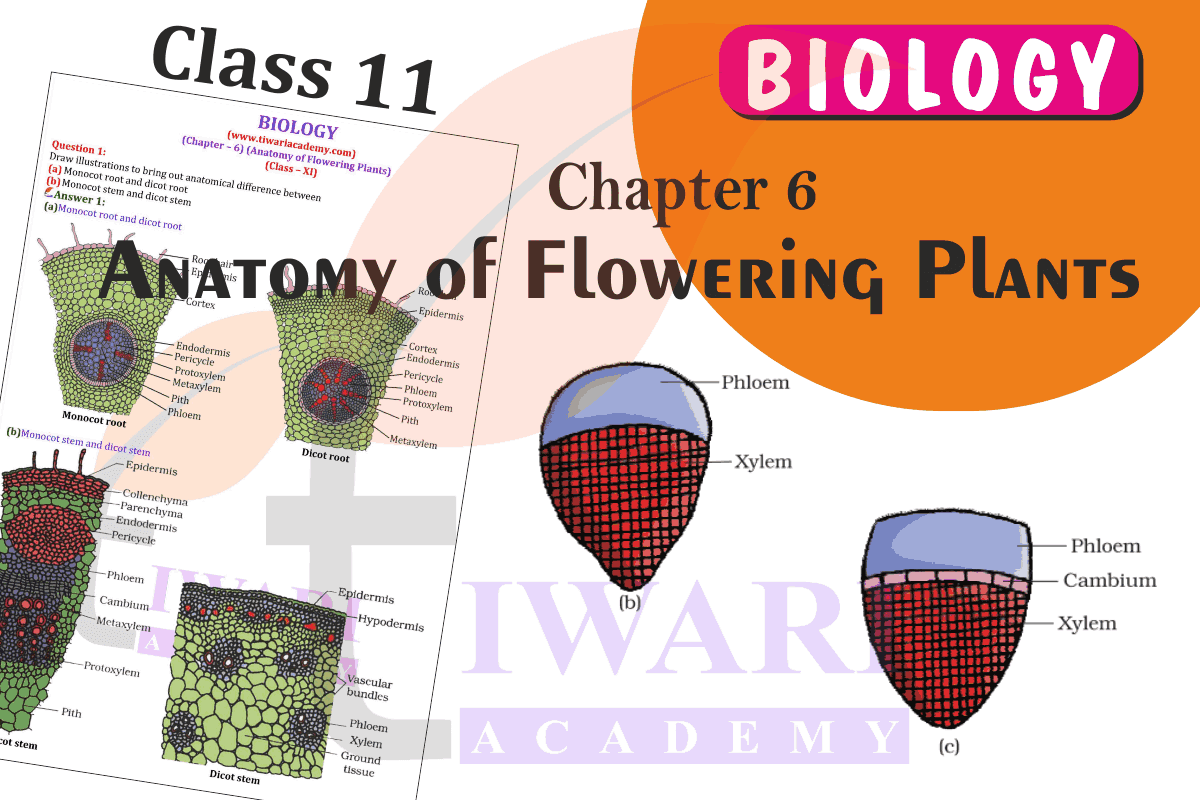
The transverse section of a plant material shows the following anatomical features, (a) the vascular bundles are conjoint, scattered and surrounded by clerenchymatous undle sheaths (b) phloem parenchyma is absent. What will you identify it as?
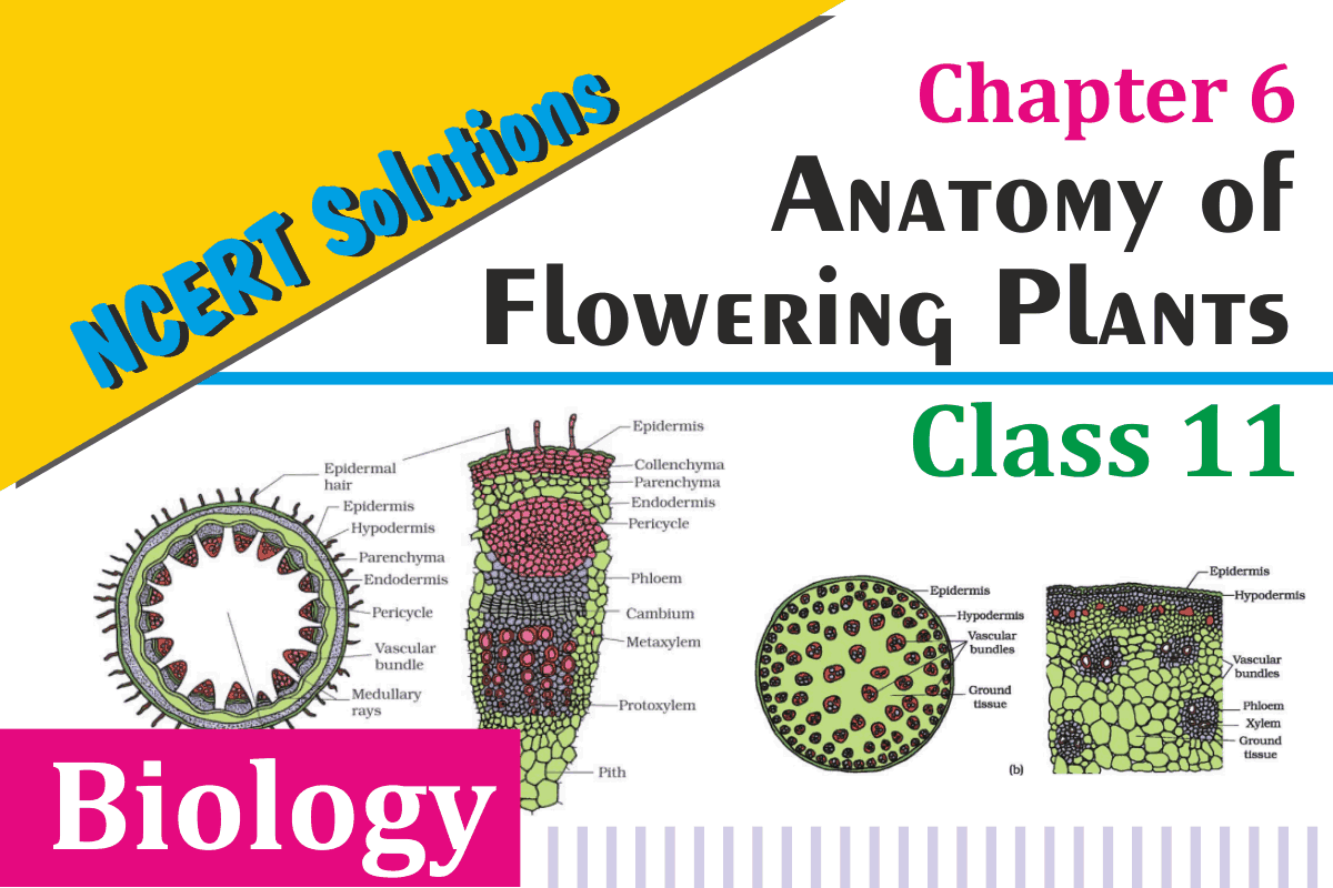
The monocot stem is characterised by conjoint, collateral, and closed vascular bundles, scattered in the ground tissue containing the parenchyma. Each vascular bundle is surrounded by sclerenchymatous bundle-sheath cells. Phloem parenchyma and medullary rays are absent in monocot stems.
Why are xylem and phloem called complex tissues?
Xylem and phloem are known as complex tissues as they are made up of more than one type of cells. These cells work in a coordinated manner, as a unit, to perform the various functions of the xylem and phloem. Xylem helps in conducting water and minerals. It also provides mechanical support to plants. It is made up of the following components: Tracheids (xylem vessels and xylem tracheids) Xylem parenchyma Xylem fibres Tracheids are elongated, thick-walled dead cells with tapering ends. Vessels are long, tubular, and cylindrical structures formed from the vessel members, with each having lignified walls and large central cavities. Both tracheids and vessels lack protoplasm. Xylem fibres consist of thick walls with an almost insignificant lumen. They help in providing mechanical support to the plant. Xylem parenchyma is made up of thin-walled parenchymatous cells that help in the storage of food materials and in the radial conduction of water. Phloem helps in conducting food materials. It is composed of: Sieve tube elements Companion cells Phloem parenchyma Phloem fibres Sieve tube elements are tube-like elongated structures associated with companion cells. The end walls of sieve tube elements are perforated to form the sieve plate. Sieve tube elements are living cells containing cytoplasm and nucleus. Companion cells are parenchymatous in nature. They help in maintaining the pressure gradient in the sieve tube elements. Phloem parenchyma helps in the storage of food and is made up of long tapering cells, with a dense cytoplasm. Phloem fibres are made up of elongated sclerenchymatous cells with thick cell walls.
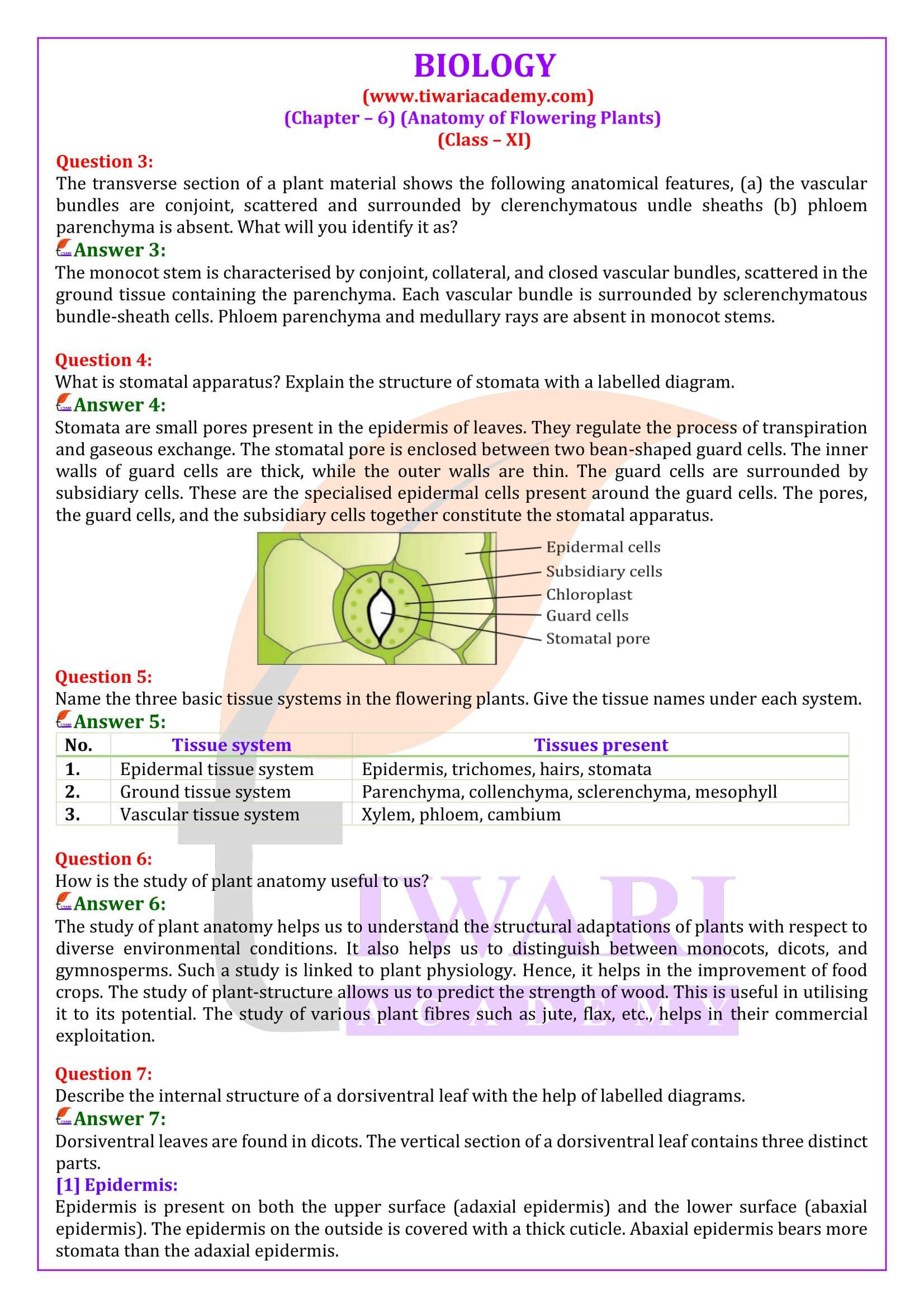
What is stomatal apparatus? Explain the structure of stomata.
Stomata are small pores present in the epidermis of leaves. They regulate the process of transpiration and gaseous exchange. The stomatal pore is enclosed between two bean-shaped guard cells. The inner walls of guard cells are thick, while the outer walls are thin. The guard cells are surrounded by subsidiary cells. These are the specialised epidermal cells present around the guard cells. The pores, the guard cells, and the subsidiary cells together constitute the stomatal apparatus.
How is the study of plant anatomy useful to us?
The study of plant anatomy helps us to understand the structural adaptations of plants with respect to diverse environmental conditions. It also helps us to distinguish between monocots, dicots, and gymnosperms. Such a study is linked to plant physiology. Hence, it helps in the improvement of food crops. The study of plant-structure allows us to predict the strength of wood. This is useful in utilising it to its potential. The study of various plant fibres such as jute, flax, etc., helps in their commercial exploitation.
What is periderm? How does periderm formation take place in dicot stem?
Periderm is composed of the phellogen, phellem, and phelloderm. During secondary growth, the outer epidermal layer and the cortical layer are broken because of the cambium. To replace them, the cells of the cortex turn meristematic, giving rise to cork cambium or phellogen. It is composed of thin-walled, narrow and rectangular cells. Phellogen cuts off cells on its either side. The cells cut off toward the outside give rise to the phellem or cork. The suberin deposits in its cell wall make it impervious to water. The inner cells give rise to the secondary cortex or phelloderm. The secondary cortex is parenchymatous.
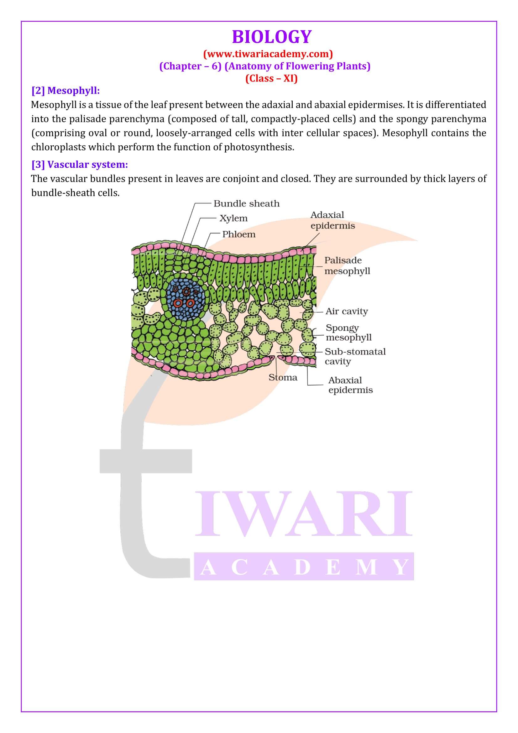
Describe the internal structure of a dorsiventral leaf.
Dorsiventral leaves are found in dicots. The vertical section of a dorsiventral leaf contains three distinct parts. [1] Epidermis: Epidermis is present on both the upper surface (adaxial epidermis) and the lower surface (abaxial epidermis). The epidermis on the outside is covered with a thick cuticle. Abaxial epidermis bears more stomata than the adaxial epidermis. [2] Mesophyll: Mesophyll is a tissue of the leaf present between the adaxial and abaxial epidermises. It is differentiated into the palisade parenchyma (composed of tall, compactly-placed cells) and the spongy parenchyma (comprising oval or round, loosely-arranged cells with inter cellular spaces). Mesophyll contains the chloroplasts which perform the function of photosynthesis. [3] Vascular system: The vascular bundles present in leaves are conjoint and closed. They are surrounded by thick layers of bundle-sheath cells.