by Tiwari Academy Team
NCERT Solutions for Class 11 Biology Chapter 8 Cell: The Unit of Life in Hindi and English Medium to Study online without download or download in PDF format free for new academic session 2026-27. CBSE NCERT Solutions of other subjects for UP board, MP board, CBSE and other boards following NCERT Books 2026-27 as a course book is available to download. Visit to Discussion Forum to share your knowledge with your classmates.
NCERT Solutions for Class 11 Biology Chapter 8
| Class: 11 | Biology |
| Chapter 8: | Cell: The Unit of Life |
| Content: | Exercises and Extra Question Answers |
| Type of Content: | PDF, Text, Videos and Images |
| Session: | Year 2026-27 |
| Medium: | Hindi and English Medium |
Class 11 Biology Chapter 8 Solutions in English
NCERT Solutions for Class 11 Biology Chapter 8 in PDF format free download is given below. NCERT Books 2026-27 and Offline Apps are available to free download for academic session 2026-27. NCERT Solutions 2026-27 are applicable for all boards who are following latest CBSE Syllabus 2026-27. Join the discussion forum to discuss your educational doubts related to all subjects.
App for Class 11 Solutions


Important Notes on Cell: The Unit of Life
1. Modification of cell envelope
2. Slime layer: Clycocalyx in form of loose sheath.
3. Capsule: Glycocalyx in form of thick and tough sheath.
4. Mesosomes: Extension of plasma membrane. These can be in the form of vesicles, tubules and lamellae.
GENETIC MATERIAL
1. It is not covered by nuclear envelope. In addition to the genomic DNA (the single chromosome/circular DNA), many bacteria have small circular self-replicating, double stranded DNA which is called as plasmid, plasmid contain genes like antibiotic resistance.
2. Cell Membrane-Singer and Nicolson (1972) gave fluid mosaic model.
According to this the quasi-fluid nature of lipid enables lateral movement of proteins within the overall bilayer; two types of proteins (Peripheral and integral proteins) with cholesterol, glycolipids and glycoproteins. Erythrocyte membrane has 52% protein and 40% lipids.
Cell Wall
1. It is non-living rigid structure which gives shape to the cell and protects cell from mechanical damage and infection, helps in cell-to-cell interaction and provides barrier to undesirable macromolecules.
2. Cell wall of algae is made of cellulose, galactans, mannans and minerals like calcium carbonate. Plant cell wall consists of cellulose, hemicellulose, pectins and proteins.
GOLGI APPARATUS
1. First observed by Camillo Golgi (in 1898)
2. Consist of cisternae stacked parallel to each other. Two faces of the organelle are convex/cis or forming face and concave/trans or maturing face.
3. Functions: Performs packaging of materials, to be delivered either to the intra-cellar targets or secreted outside the cell. Important site of formation of glycoproteins and glycolipids.
Important Questions on 11th Biology Chapter 8
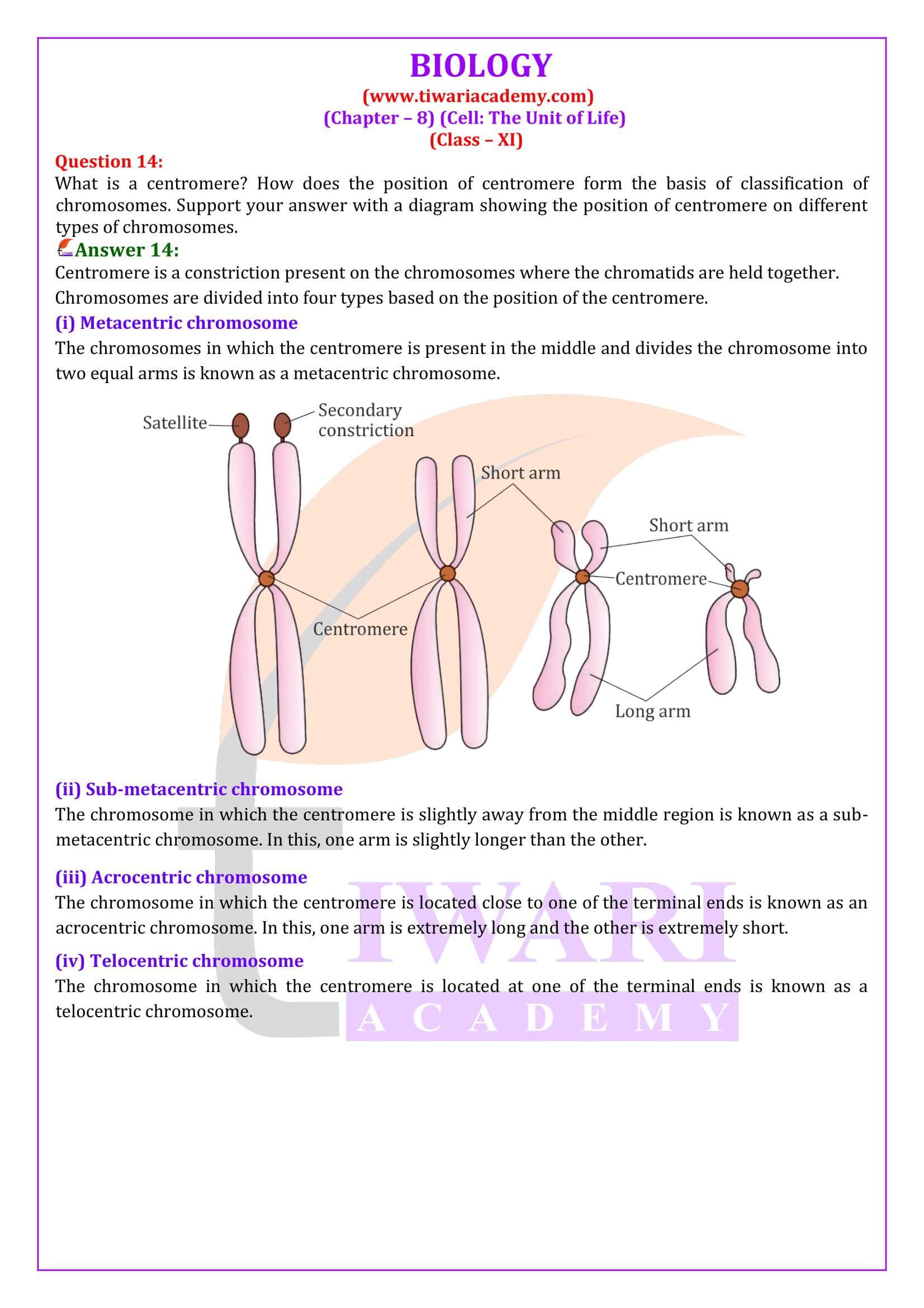
What is a mesosome in a prokaryotic cell? Mention the functions that it performs.
Mesosome is a convoluted membranous structure formed in a prokaryotic cell by the invagination of the plasma membrane. Its functions are as follows: These extensions help in the synthesis of the cell wall, replication of DNA. They also help in the equal distribution of chromosomes into the daughter cells. It also increases the surface area of the plasma membrane to carry out various enzymatic activities. It helps in secretion processes as well as in bacterial respiration.
How do neutral solutes move across the plasma membrane? Can the polar molecules also move across it in the same way? If not, then how are these transported across the membrane?
Plasma membrane is the outermost covering of the cell that separates it from the environment. It regulates the movement of substances into the cell and out from it. It allows the entry of only some substances and prevents the movement of other materials. Hence, the membrane is selectively-permeable. Movement of neutral solutes across the cell membrane – Neutral molecules move across the plasma membrane by simple passive diffusion. Diffusion is the movement of molecules from a region of higher concentration to a region of lower concentration. Movement of polar molecules across the cell membrane – The cell membrane is made up of a phospholipid bilayer and proteins. The movement of polar molecules across the non-polar lipid bilayer requires carrier-proteins. Carrier-proteins are integral protein particles having certain affinity for specific solutes. As a result, they facilitate the transport of molecules across the membrane.
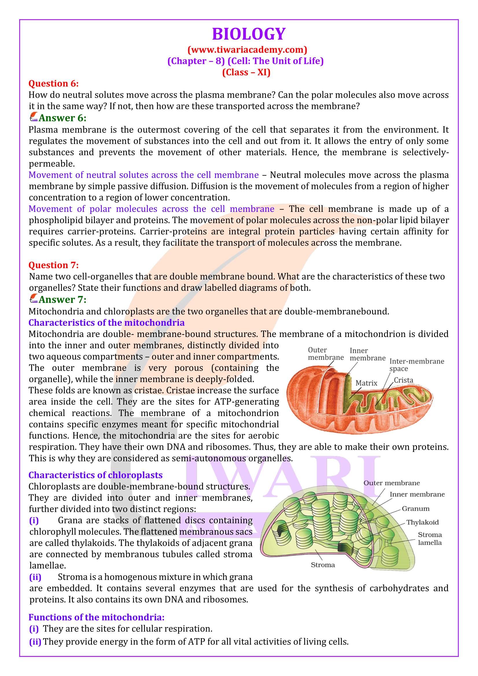
Name two cell-organelles that are double membrane bound. What are the characteristics of these two organelles? State their functions.
Mitochondria and chloroplasts are the two organelles that are double-membranebound. Characteristics of the mitochondria Mitochondria are double- membrane-bound structures. The membrane of a mitochondrion is divided into the inner and outer membranes, distinctly divided into two aqueous compartments – outer and inner compartments. The outer membrane is very porous (containing the organelle), while the inner membrane is deeply-folded. These folds are known as cristae. Cristae increase the surface area inside the cell. They are the sites for ATP-generating chemical reactions. The membrane of a mitochondrion contains specific enzymes meant for specific mitochondrial functions. Hence, the mitochondria are the sites for aerobic respiration. They have their own DNA and ribosomes. Thus, they are able to make their own proteins. This is why they are considered as semi-autonomous organelles Characteristics of chloroplasts Chloroplasts are double-membrane-bound structures. They are divided into outer and inner membranes, further divided into two distinct regions: (i) Grana are stacks of flattened discs containing chlorophyll molecules. The flattened membranous sacs are called thylakoids. The thylakoids of adjacent grana are connected by membranous tubules called stroma lamellae. (ii) Stroma is a homogenous mixture in which grana are embedded. It contains several enzymes that are used for the synthesis of carbohydrates and proteins. It also contains its own DNA and ribosomes. Functions of the mitochondria: (i) They are the sites for cellular respiration. (ii) They provide energy in the form of ATP for all vital activities of living cells. (iii) They have their own DNA and ribosomes. Hence, they are regarded as semiautonomous organelles. (iv) They have several enzymes, intermediately required for the synthesis of various chemicals such as fatty acids, steroids, and amino acids. Functions of chloroplasts: (i) They trap solar energy and utilise it for manufacturing food for plants. Hence, they are involved in the process of photosynthesis. (ii) They contain the enzymes required for the synthesis of carbohydrates and proteins.
What are the characteristics of prokaryotic cells?
Prokaryotic cell is a unicellular organism lacking membrane-bound organelles. The characteristics of prokaryotic cells are as follows: Most of them are unicellular. They are generally small in size. The size of a prokaryotic cell varies from 0.5 – 5 µm. The nuclear region of a prokaryotic cell is poorly-defined because of the absence of a nuclear membrane. Hence, a prokaryotic cell lacks a true nucleus. The genetic materials of prokaryotic cells are naked. They contain single, circular chromosomes. In addition to the genomic DNA, they have a small, circular plasmid DNA. They have specialised membranous structures called mesosomes. Mesosomes are formed by the invagination of the cell membrane. These extensions help in the synthesis of the cell wall, replication of DNA. They also help in the equal distribution of chromosomes into the daughter cells. Membrane-bound cell organelles such as mitochondria, plastids, and endoplasmic reticulum are absent from a prokaryotic cell. Most prokaryotic cells contain a three-layered structure – outermost glycocalyx, middle cell wall, and the innermost plasma membrane. This structure acts as a protective unit. Examples of prokaryotic cells include blue green algae, bacteria, etc.
Multicellular organisms have division of labour. Explain.
Multicellular organisms are made up of millions and trillions of cells. All these cells perform specific functions. All the cells specialised for performing similar functions are grouped together as tissues in the body. Hence, a particular function is carried out by a group of cells at a definite place in the body. Similarly, different functions are carried out by different groups of cells in an organism. This is known as division of labour in multicellular organisms.
Cell is the basic unit of life. Discuss in brief.
Cells are the basic units of life capable of doing all the required biochemical processes that a normal cell has to do in order to live. The basic needs for the survival of all living organisms are the same. All living organisms need to respire, digest food for obtaining energy, and get rid of metabolic wastes. Cells are capable of performing all the metabolic functions of the body. Hence, cells are called the functional units of life.
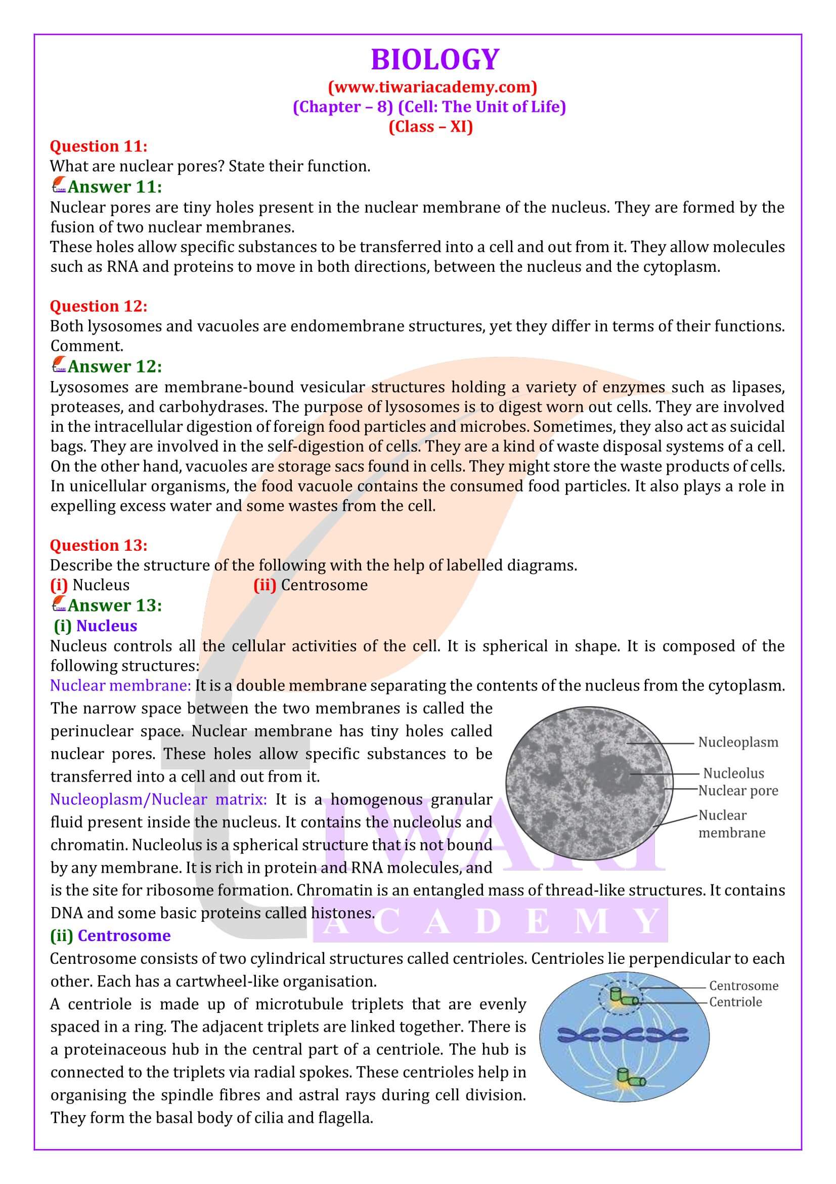
What are nuclear pores? State their function.
Nuclear pores are tiny holes present in the nuclear membrane of the nucleus. They are formed by the fusion of two nuclear membranes. These holes allow specific substances to be transferred into a cell and out from it. They allow molecules such as RNA and proteins to move in both directions, between the nucleus and the cytoplasm.
Both lysosomes and vacuoles are endomembrane structures, yet they differ in terms of their functions. Comment.
Lysosomes are membrane-bound vesicular structures holding a variety of enzymes such as lipases, proteases, and carbohydrases. The purpose of lysosomes is to digest worn out cells. They are involved in the intracellular digestion of foreign food particles and microbes. Sometimes, they also act as suicidal bags. They are involved in the self digestion of cells. They are a kind of waste disposal systems of a cell. On the other hand, vacuoles are storage sacs found in cells. They might store the waste products of cells. In unicellular organisms, the food vacuole contains the consumed food particles. It also plays a role in expelling excess water and some wastes from the cell.
Lysosomes
1. Membrane bound vesicular structures formed by the process of packaging in the golgi apparatus. Contain hydrolysing enzymes (lipases, proteases, carbohydrates) which are active in acidic pH. Also called Suicidal Bag.
2. Function: Intracellular digestion.