by Tiwari Academy Team
NCERT Solutions for Class 11 Biology Chapter 17 Locomotion and Movement in Hindi and English to Study online as well as to download in PDF format free for academic session 2026-27. Download NCERT Solutions of Chemistry, Maths, Business Studies and other subjects also. Join Discussion Forum and share your views in the world of knowledge.
NCERT Solutions for Class 11 Biology Chapter 17
Class 11 Biology Chapter 17 Locomotion and Movement Solutions
| Class: 11 | Biology |
| Chapter 17: | Locomotion and Movement |
| Content: | Study Material and Exercises Solutions |
| Mode of Content: | PDF, Images and Videos Format |
| Medium used: | English, Hindi |
| Academic Session: | Year 2026-27 |
Class 11 Biology Chapter 17 Solutions in English
NCERT Solutions for Class 11 Biology Chapter 17 in PDF format free download for new session 2026-27. Join the discussion forum to ask your doubts and discuss your questions with our experts. Download NCERT Books 2026-27 based on latest CBSE Syllabus for all boards who are using NCERT Books are course books. If you have any query related to NIOS Board, ask through discussion forum.
Download App for 11th Class


Important Notes on Locomotion and Movement
TYPES OF MUSCLES
1. Skeletal muscles or striated muscles: These involved in locomotion and change of body postures. These are also known as voluntary muscles.
2. Visceral muscles or smooth muscles: These are located in inner wall of hollow visceral organ, smooth in appearance and their activity are not under control of nervous system. They are called involuntary muscles.
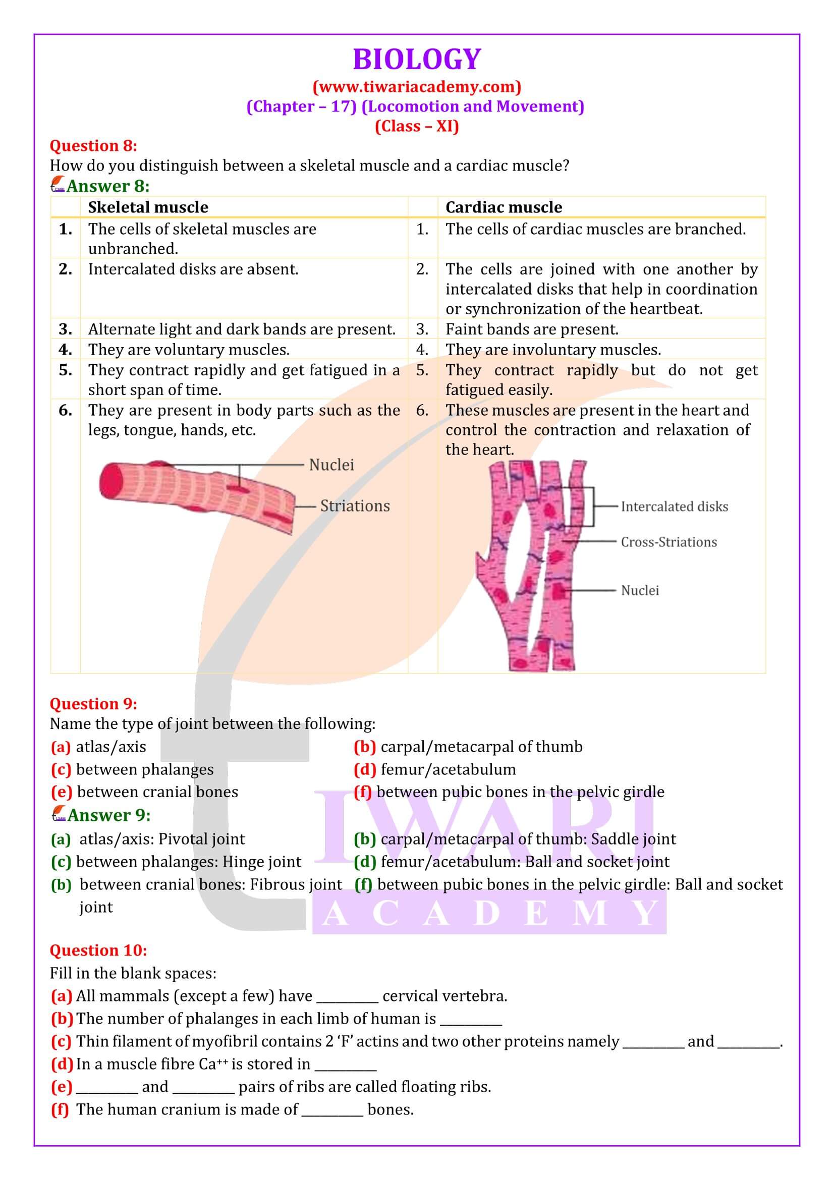
3. Cardiac muscles: The muscles of heart, involuntary in nature, striated and branched, These are uninucleated.
White muscle fibres
1. These are pale or whitish due to presence of less content of myoglobin.
2. These contain fewer mitochondria
3. Sarcoplasmic reticulum is more/high
4. During strenuous exercise, lactic acid accumulates in large quantity so muscle fatigues
Red muscle fibres
1. These are red in colour due to presence of high content of myoglobin.
2. These contain plenty of mitochondria.
3. Sarcoplasmic reticulum is less in these fibres.
4. Show slow but sustained contractions for longer periods.
TYPES OF MOVEMENT
1. Amoeboid movement: These movements takes place in phagocytes where leucocytes and macrophages migrate through tissue. It is affected by pseudopodia formed by the streaming of protoplasm (as in amoeba)
2. Ciliary movement: These movement occurs in internal organs which are lined by ciliary epithelium.
3. Muscular Movement: This movements involve the muscle fibres, which have the ability to contract and relax.
Important Questions on 11th Biology Chapter 17
Define sliding filament theory of muscle contraction.
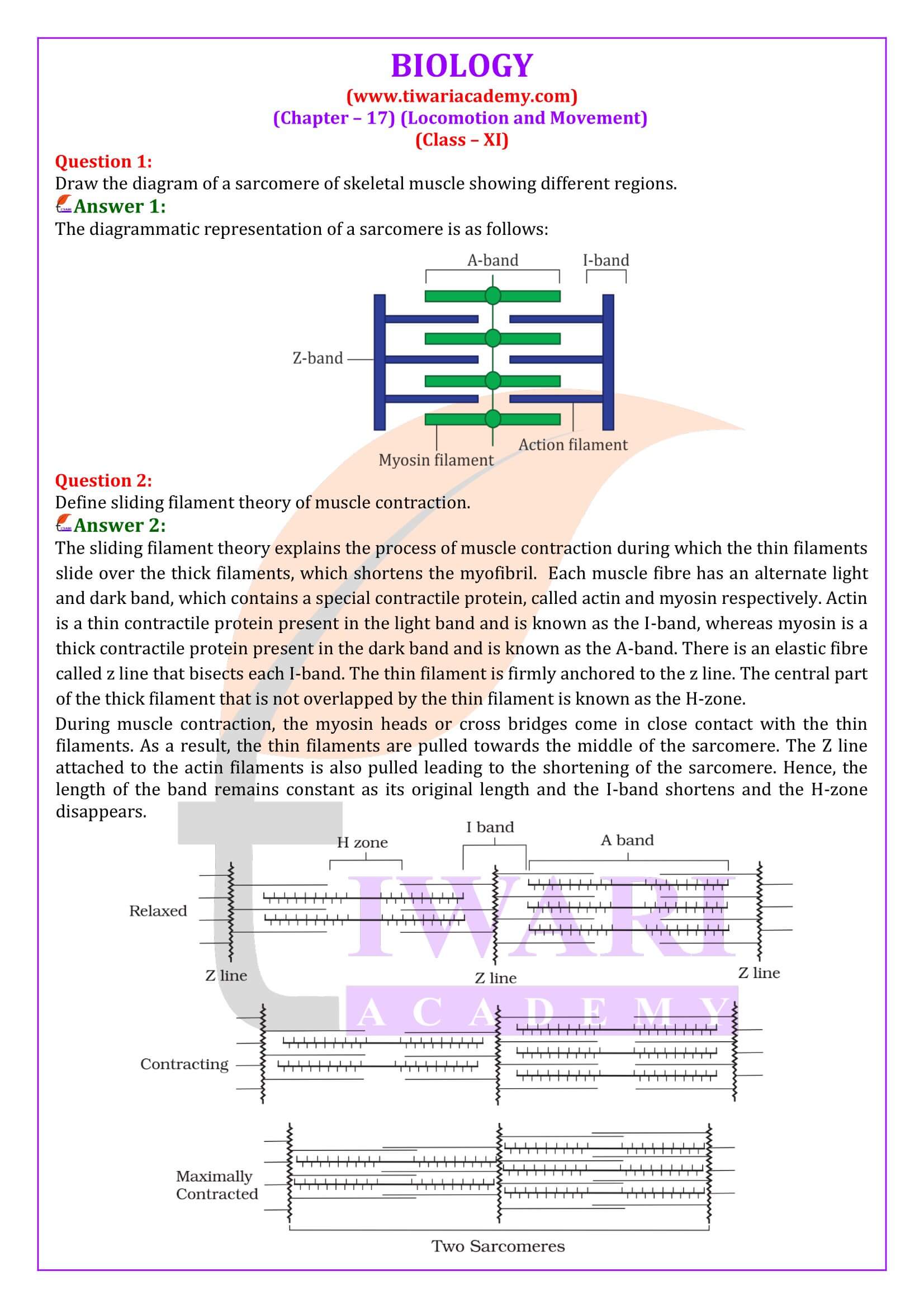
The sliding filament theory explains the process of muscle contraction during which the thin filaments slide over the thick filaments, which shortens the myofibril. Each muscle fibre has an alternate light and dark band, which contains a special contractile protein, called actin and myosin respectively. Actin is a thin contractile protein present in the light band and is known as the I-band, whereas myosin is a thick contractile protein present in the dark band and is known as the A-band. There is an elastic fibre called z line that bisects each I-band. The thin filament is firmly anchored to the z line. The central part of the thick filament that is not overlapped by the thin filament is known as the H-zone. During muscle contraction, the myosin heads or cross bridges come in close contact with the thin filaments. As a result, the thin filaments are pulled towards the middle of the sarcomere. The Z line attached to the actin filaments is also pulled leading to the shortening of the sarcomere. Hence, the length of the band remains constant as its original length and the I-band shortens and the H-zone disappears.
Describe the important steps in muscle contraction.
During skeletal muscle contraction, the thick filament slides over the thin filament by a repeated binding and releases myosin along the filament. This whole process occurs in a sequential manner. Step 1: Muscle contraction is initiated by signals that travel along the axon and reach the neuromuscular junction or motor end plate. Neuromuscular junction is a junction between a neuron and the sarcolemma of the muscle fibre. As a result, Acetylcholine (a neurotransmitter) is released into the synaptic cleft by generating an action potential in sarcolemma. Step 2: The generation of this action potential releases calcium ions from the sarcoplasmic reticulum in the sarcoplasm. Step 3: The increased calcium ions in the sarcoplasm leads to the activation of actin sites. Calcium ions bind to the troponin on actin filaments and remove the tropomyosin, wrapped around actin filaments. Hence, active actin sites are exposed and this allows myosin heads to attach to this site. Step 4: In this stage, the myosin head attaches to the exposed site of actin and forms cross bridges by utilizing energy from ATP hydrolysis. The actin filaments are pulled. As a result, the H-zone reduces. It is at this stage that the contraction of the muscle occurs. Step 5: After muscle contraction, the myosin head pulls the actin filament and releases ADP along with inorganic phosphate. ATP molecules bind and detach myosin and the cross bridges are broken. Stage 6: This process of formation and breaking down of cross bridges continues until there is a drop in the stimulus, which causes an increase in calcium. As a result, the concentration of calcium ions decreases, thereby masking the actin filaments and leading to muscle relaxation.
What are the different types of movements exhibited by the cells of human body?
Movement is a characteristic feature of living organisms. The different types of movement exhibited by cells of the human body are: Amoeboid movement: Leucocytes present in the blood show amoeboid movement. During tissue damage, these blood cells move from the circulatory system towards the injury site to initiate an immune response. Ciliary movement: Reproductive cells such as sperms and ova show ciliary movement. The passage of ova through the fallopian tube towards the uterus is facilitated by this movement. Muscular movement: Muscle cells show muscular movement.
Structure of Actin and Myosin Filament
1. Actin filament: An actin filament is made of two ‘F’ actins which are helically wound to each other. Two filaments of tropo myosin protein also run close to ‘F’ actins throughout its length. A complex protein Troponin is distributed at regular intervals on tropomyosin which mask the actin binding site for myosin.
2. Myosin filament: Each myosin filament is a polymer of meromyosin. Each meromyosin has two components a globular head with a short arm and a tail. Head is made of heavy meromyosin while tail is made of light meromyosin. The head with its short arm project outward at regular distance and angle from each other and is known as cross arm. The head has an active site for actin and binding site for ATP.